|
所属 |
医学部 医学科 機能制御学講座血管動態生化学分野 |
|
職名 |
教授 |
|
研究室住所 |
宮崎県宮崎市清武町木原5200 |
|
研究室電話番号 |
0985-85-0985 |
|
連絡先 |
|
|
ホームページ |
|
|
外部リンク |
|
|
関連SDGs |
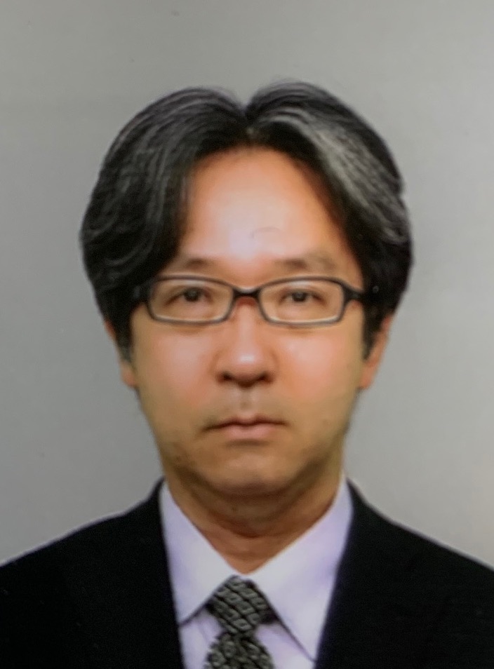
西山 功一 (ニシヤマ コウイチ)
NISHIYAMA Koichi
|
|
|
研究分野 【 表示 / 非表示 】
-
ライフサイエンス / 生物物理学
-
ライフサイエンス / 生体医工学
-
ライフサイエンス / 細胞生物学
-
ライフサイエンス / 分子生物学
-
ライフサイエンス / 循環器内科学
学内職務経歴 【 表示 / 非表示 】
-
宮崎大学 医学部 医学科 機能制御学講座血管動態生化学分野 教授
2021年06月 - 継続中
-
宮崎大学 医学部 医学科 機能制御学講座腫瘍生化学分野 教授
2021年04月 - 2021年05月
学外略歴 【 表示 / 非表示 】
-
熊本大学 附属病院 講師
2014年4月 - 2016年3月
-
熊本大学 国際先端医学研究機構 主任研究員
2014年 - 2021年3月
-
東京大学 大学院医学系研究科 助教
2006年4月 - 2014年3月
-
東京大学 大学院医学系研究科 研究員
2005年4月 - 2006年3月
論文 【 表示 / 非表示 】
-
Mechanical loading of intraluminal pressure mediates wound angiogenesis by regulating the TOCA family of F-BAR proteins 査読あり 国際共著 国際誌
Yuge S., Nishiyama K., Arima Y., Hanada Y., Oguri-Nakamura E., Hanada S., Ishii T., Wakayama Y., Hasegawa U., Tsujita K., Yokokawa R., Miura T., Itoh T., Tsujita K., Mochizuki N., Fukuhara S.
Nature Communications 13 ( 1 ) 2594 - 2594 2022年12月
担当区分:筆頭著者, 責任著者 記述言語:英語 掲載種別:研究論文(学術雑誌) 出版者・発行元:Nature Communications
Angiogenesis is regulated in coordinated fashion by chemical and mechanical cues acting on endothelial cells (ECs). However, the mechanobiological mechanisms of angiogenesis remain unknown. Herein, we demonstrate a crucial role of blood flow-driven intraluminal pressure (IP) in regulating wound angiogenesis. During wound angiogenesis, blood flow-driven IP loading inhibits elongation of injured blood vessels located at sites upstream from blood flow, while downstream injured vessels actively elongate. In downstream injured vessels, F-BAR proteins, TOCA1 and CIP4, localize at leading edge of ECs to promote N-WASP-dependent Arp2/3 complex-mediated actin polymerization and front-rear polarization for vessel elongation. In contrast, IP loading expands upstream injured vessels and stretches ECs, preventing leading edge localization of TOCA1 and CIP4 to inhibit directed EC migration and vessel elongation. These data indicate that the TOCA family of F-BAR proteins are key actin regulatory proteins required for directed EC migration and sense mechanical cell stretching to regulate wound angiogenesis.
-
Biomechanical control of vascular morphogenesis by the surrounding stiffness 査読あり
花田 保之, 小河 穂波, 西山 功一
Nature Communications 16 6788 2025年7月
記述言語:英語 掲載種別:研究論文(学術雑誌) 出版者・発行元:Springer Nature
Sprouting angiogenesis is a form of morphogenesis which expands vascular networks from preexisting networks. However, the precise mechanism governing efficient branch elongation driven by directional movement of endothelial cells (ECs), while the lumen develops under the influence of blood inflow, remains unknown. Herein, we show perivascular stiffening to be a major factor that integrates branch elongation and lumen development. The lumen expansion seen during lumen development inhibits directional EC movement driving branch elongation. This process is counter-regulated by the presence of pericytes, which induces perivascular stiffening by promoting the deposition of EC-derived collagen-IV (Col-IV) on the vascular basement membrane (VBM), thereby preventing excessive lumen expansion. Furthermore, inhibition of forward directional movement of the tip EC during lumen development is associated with decreased localization of the F-BAR proteins and Arp2/3 complexes at the leading front. Our results demonstrate how ECs elongate branches, while the lumen develops, by properly building the surrounding physical environment in coordination with pericytes during angiogenesis.
-
Biomechanical control of vascular morphogenesis by the surrounding stiffness. 査読あり 国際誌
Hanada Y, Halder S, Arima Y, Haruta M, Ogoh H, Ogura S, Shiraki Y, Nakano S, Ozeki Y, Fukuhara S, Uemura A, Murohara T, Nishiyama K
Nature communications 16 ( 1 ) 6788 - 6788 2025年7月
担当区分:責任著者 記述言語:英語 掲載種別:研究論文(学術雑誌)
Sprouting angiogenesis is a form of morphogenesis which expands vascular networks from preexisting networks. However, the precise mechanism governing efficient branch elongation driven by directional movement of endothelial cells (ECs), while the lumen develops under the influence of blood inflow, remains unknown. Herein, we show perivascular stiffening to be a major factor that integrates branch elongation and lumen development. The lumen expansion seen during lumen development inhibits directional EC movement driving branch elongation. This process is counter-regulated by the presence of pericytes, which induces perivascular stiffening by promoting the deposition of EC-derived collagen-IV (Col-IV) on the vascular basement membrane (VBM), thereby preventing excessive lumen expansion. Furthermore, inhibition of forward directional movement of the tip EC during lumen development is associated with decreased localization of the F-BAR proteins and Arp2/3 complexes at the leading front. Our results demonstrate how ECs elongate branches, while the lumen develops, by properly building the surrounding physical environment in coordination with pericytes during angiogenesis.
-
第1土曜特集 "かたちづくり" を制御する分子メカニズム 形態形成と多細胞動態 血管新生における血流による物理的力の役割 招待あり
花田 保之, 西山 功一
医学のあゆみ 290 ( 1 ) 42 - 46 2024年7月
-
第5土曜特集 血管・リンパ管研究の最前線と治療への展開 血管研究のフロンティア 再構成解析系を駆使した血流による血管新生の生体力学機序の解明 招待あり
西山 功一
医学のあゆみ 289 ( 13 ) 1093 - 1098 2024年6月
MISC 【 表示 / 非表示 】
-
血流に起因する内腔圧による創傷治癒過程の血管新生の新たな制御機構
福原茂朋, 弓削進弥, 西山功一, 有馬勇一郎, 花田保之, 花田三四郎, 石井智裕, 若山勇紀, 辻田和也, 横川隆司, 三浦岳, 望月直樹
日本生化学会大会(Web) 93rd 2020年
担当区分:筆頭著者 記述言語:日本語 掲載種別:速報,短報,研究ノート等(学術雑誌)
-
花田 保之, 西山 功一
医学のあゆみ 290 ( 1 ) 42 - 46 2024年7月
記述言語:日本語 掲載種別:記事・総説・解説・論説等(学術雑誌) 出版者・発行元:東京 : 医歯薬出版
その他リンク: https://ndlsearch.ndl.go.jp/books/R000000004-I033576985
-
創傷治癒過程の血管新生における内腔圧の新たな役割の解明 招待あり
福原茂朋, 弓削進弥, 有馬勇一郎, 花田保之, 花田三四郎, 石井智裕, 若山勇紀, 横川隆司, 三浦岳, 望月直樹, 西山功一
脈管学(Web) 60 ( supplement ) 2020年
記述言語:日本語 掲載種別:研究発表ペーパー・要旨(全国大会,その他学術会議)
-
内腔圧の機械的刺激により制御される創傷治癒での血管新生
弓削進弥, 西山功一, 有馬勇一郎, 花田保之, 花田三四郎, 石井智裕, 若山勇紀, 辻田和也, 横川隆司, 三浦岳, 望月直樹, 福原茂朋
日本生化学会大会(Web) 93rd 2020年
記述言語:日本語 掲載種別:速報,短報,研究ノート等(学術雑誌)
科研費(文科省・学振・厚労省)獲得実績 【 表示 / 非表示 】
-
ペリサイトの血流作用統合による血管新生の生体力学的制御機構の解明
研究課題/領域番号:24K03267 2024年04月 - 2027年03月
独立行政法人日本学術振興会 科学研究費基金 基盤研究(B)
西山 功一, 植村 明嘉
担当区分:研究代表者
-
オルガノイド血管化・灌流によるヒト腎糸球体構造・機能の生体外再現法の開発
研究課題/領域番号:24K22384 2024年04月 - 2027年03月
独立行政法人日本学術振興会 科学研究費基金 挑戦的研究(萌芽)
西山 功一, 田中 悦子
担当区分:研究代表者
-
血流とペリオサイトの協奏による血管新生メカノバイオロジー機構
研究課題/領域番号:19H04446 2021年04月 - 2024年03月
独立行政法人日本学術振興会 科学研究費助成事業 基盤研究(B) 基盤研究(B)
西山 功一, 梅本 晃正, 植村 明嘉
担当区分:研究代表者
本研究では、血流による血管内腔圧・壁伸展刺激が内皮細胞により感知・伝達され、血管新生が抑制されるメカニズムを明らかにし、さらに、ペリサイトがその内腔圧・壁伸展刺激を調節し血管新生を促進的に制御するメカニズムを明らかにすることで、『血流とペリサイトの協奏による血管新生メカノバイオロジー機構』という全く新しい血管新生メカニズムの概念を提唱することを目的とした。
昨年度までのオンチップモデル解析から、内皮細胞が血管内腔圧上昇に伴う細胞膜の伸展刺激を受けると血管伸長が抑制されることを見出し、それは、内皮細胞の前後極性と方向性運動の障害に起因していることがわかった。本年度においては、さらに、内皮細胞膜が伸展を受けることで膜結合型BARタンパクFNBP1LおよびCIP4が膜から解離し、Arp2/3複合体の先導端局在とアクチン重合の失敗により内皮細胞の前後極性形成が失われ、方向性運動が障害されるというメカノバイオロジー機構が明らかとなった。また、両遺伝子のin vivoでの重要性を今後検討していくために、共同研究者と共に、FNBP1LおよびCIP4のコンベンショナルノックアウトマウスの立ち上げを行った。
一方、昨年度までのオンチップとマウス網膜血管新生の解析にて、ペリサイトは血管の伸長を促進し、血管径を小さく保つことがわかった。本年度においては、オンチップ血管新生におけるタイムラプス観察等から、ペリサイトは、血管径の過度な拡大を抑えて血管壁にかかる伸展張力を制御し、内皮細胞の方向性運動の効率性を保つことで、枝の伸長を促進する生体力学機構が示唆されてきた。さらに、ペリサイトの存在下では、血管壁周囲の細胞外基質の硬度が上がり、血管径の過度な拡大が抑えられているしくみが明らかとなった。細胞外基質硬度が上がるメカニズムに関しては、現在検討中である。 -
血管化灌流によるヒト腎糸球体構造・機能の生体外再現と応用
研究課題/領域番号:21K19487 2021年04月 - 2023年03月
独立行政法人日本学術振興会 科学研究費助成事業 挑戦的研究(萌芽) 挑戦的研究(萌芽)
西山 功一
担当区分:研究代表者
慢性腎臓病は、様々な疾患死亡のリスク因子であり、国民の生命と健康の維持そして逼迫する我が国の医療経済のため克服すべき重大な課題である。本研究では、ヒト腎オルガノイドを血管化・灌流し高次糸球体構造と濾過機能を生体外で再現する、これまで実現されていない培養システムの開発、さらに、同システムを用いて、血管化・灌流を受けて高次糸球体構造が形成され機能を獲得し維持されるしくみを明らかにすることを目指した。
令和3年度において、まず、ヒトiPS由来腎臓前駆細胞のスフィアから腎臓オルガノイドを誘導する方法を、マウス胎仔神経管との共培養により発生誘導をかける方法から、CHIRによる分化誘導法に変更し、その方法論的確立を行った。また、これまで、気液界面培養にて、腎糸球体発生とオルガノイド内血管網形成を進めた後に回転浮遊培養に移行すると、糸球体血管化が誘導される予備的知見を得ていた。したがって、その最適な移行タイミング、ならびに、内皮細胞にて血管灌流が可能な自己組織化血管網を構築した微小流体デバイス(血管灌流チップ)にオルガノイドを移植するタイミングの最適条件を検討した。その結果、CHIRで発生誘導後のオルガノイドを、気液界面培養にて7日程度さらに発生を進め、回転培養でさらに1週間培養すると、最も糸球体内に血管が侵入することがわかった。しかし、その後、同オルガノイドを血管灌流チップに導入し、血管化・灌流培養を行ったが、灌流により糸球体内への血管の侵入は促進されるものの、灌流培養7日後においても、糸球体濾過装置を形成する血管化され成熟した糸球体を誘導できる条件は見出せなかった。 -
ペリサイト消失網膜における炎症と線維化の細胞・分子機構の解明
研究課題/領域番号:19H03437 2019年04月 - 2022年03月
日本学術振興会 科学研究費助成事業 基盤研究(B) 基盤研究(B)
植村 明嘉, 西山 功一
担当区分:研究分担者
抗PDGFRβ抗体を腹腔内に単回投与して、ペリサイトを消失させた新生仔マウス網膜では、①内皮細胞の炎症反応、②内在性ミクログリアの活性化と骨髄由来マクロファージの浸潤、③血管透過性の亢進、④網膜剥離の発症、⑤活性化型ミクログリアの網膜下への移動、⑥急性炎症から慢性炎症への移行、⑦網膜下の線維化が、順に進行する。こうした過程で、抗CSF1R抗体を投与してミクログリアを消失させると、線維化が抑制されることから、網膜下に移行した活性化型ミクログリアが線維化を誘導することが明らかとなった。さらに単細胞RNAseq解析により、線維化誘導ミクログリアがM2極性化していることが明らかとなった。
寄附金・講座・研究部門 【 表示 / 非表示 】
-
機能制御学講座血管動態生化学分野研究奨学金(公益財団法人ノバルティス科学振興財団)
2025年03月
-
機能制御学講座血管動態生化学分野研究奨学金(第一三共生命科学研究振興財団)
2024年10月
-
機能制御学講座血管動態生化学分野研究奨学金(ジョンソン・エンドジョンソン株式会社)
2024年10月
-
機能制御学講座血管動態生化学分野研究奨学金
2024年09月
-
血管動態制科学分野研究奨学金(中谷医工計測技術振興財団)
寄附者名称:公益財団法人 中谷医工計測技術振興財団 2024年02月
研究・技術シーズ 【 表示 / 非表示 】
-
血管新生のしくみの解明と医療応用

物流ネットワークとしての血管形成とその破綻による病気の理解と制御
血流がある生命現象の試験管内モデルの開発と医学・薬学への応用ホームページ: 医学部機能制御学講座血管動態制御学分野
技術相談に応じられる関連分野:1.血管新生の解析全般 2.タイムラプスイメージングによる分子・細胞動態の定量的解析 3.ホールマウント免疫染色と画像解析、定量化 4.血管チップを使ったオルガノイド、組織の培養 5.血管チップを使った力学刺激の評価、医療応用
メッセージ:医学・生物学にとらわれない視点で、多くの共同研究者とともに学際的な研究を推進しています。興味を持たれた方はお気軽にお声掛けください。


